Diffusion Spectrum Imaging (DSI)
Diffusion Spectrum Imaging (DSI) | In vivo Magnetic Resonance Spectroscopy | Stroke Imaging | Cerebral Blood Flow Imaging| Diffusion Tensor Imaging
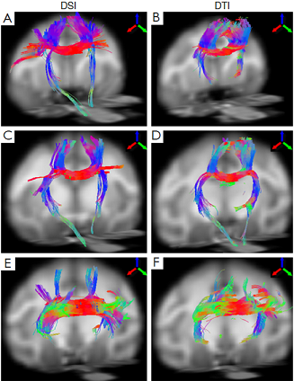
Conventional diffusion tensor imaging (DTI) cannot directly image multiple fiber orientations within a single voxel. In comparison, diffusion spectrum imaging (DSI) can detect the multiple fiber orientation within a voxel and allows for delineating crossing and touching fibers in the brain. As demonstrated below, transcallosal fiber tracts of adult macaque brains are examined with DSI and compared with those from a conventional DTI protocol. The results demonstrate that DSI can reveal the transcallosal fiber bundles much more extensively than the conventional DTI, suggesting that DSI can provide a feasible and robust approach for characterizing the fiber pathways and brain connectivity in NHPs.
 |
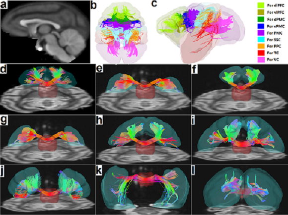 |
Figure 2. Demonstration of the transcallosal fiber tracts for various cortical areas in an adult monkey brain using ex vivo DSI on a Bruker 7T. The fiber tracking results are from a fixed adult macaque brain (~10 years old), overlaid on the DSI B0 images. (Meng and Zhang, Brain Connectivity, 2014 (in press). PMID: 25389564)
|
Directions | Contact Us | ©2022 EPC Imaging Center