Stroke Imaging
Diffusion Spectrum Imaging (DSI) | In vivo Magnetic Resonance Spectroscopy | Stroke Imaging | Cerebral Blood Flow Imaging| Diffusion Tensor Imaging
MRI of acute stroke
MRI is the only imaging technique that can detect stroke injury as early as few minutes after insult. Multi-parameter MRI is increasingly used in the diagnosis of stroke. We have developed multiple MRI techniques including quantitative structural, perfusion, diffusion and functional MRI to image stroke injury with/without treatment.
MRI for monkey brains with stroke
 |
 |
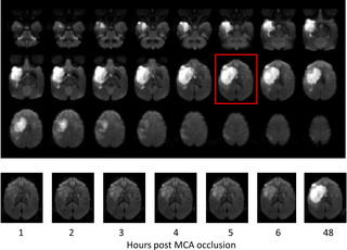
| Fig 1. Diffusion weighted images illustrate stroke lesion evolution in an adult rhesus monkey brain. X Zhang et al, PLOS one, 2015. |
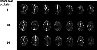
Fig 2. Perfusion MRI illustrates temporal perfusion changes in a monkey brain after permanent MCA occlusion. X Zhang et al, PLOS one, 2015 |
MRI for rat and mouse brains with stroke
 |
 |
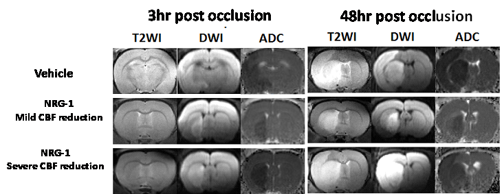
| Fig 3. Diffusion weighted images (DWI), T2-weighted images (T2WI), ADC maps illustrate stroke lesion evolution in a rat brain with/without treatment. Wang et al, JNS, 2015. |
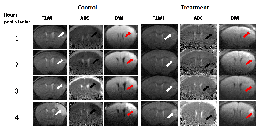
Fig 4. Diffusion weighted images (DWI), T2-weighted images (T2WI), ADC maps illustrate stroke lesion evolution in a mouse brain with/without treatment. Wang et al, ISMRM, 2015 |
Directions | Contact Us | ©2022 EPC Imaging Center